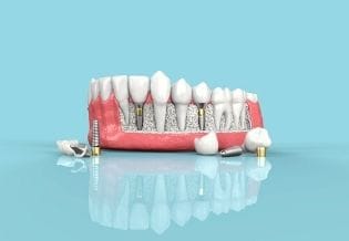
Maxillary Sinus Lift Using the Lateral Window Technique with Hydroxyapatite and Beta-Tricalcium Phosphate (β-TCP) Graft
Abstract
This narrative literature review investigates the clinical feasibility of maxillary sinus lift using the lateral window (or traumatic) technique, employing a grafting material composed of hydroxyapatite associated with beta-tricalcium phosphate. The study is based on the premise that bone resorption and sinus pneumatization—common in edentulous posterior maxillae—pose a challenge to achieving primary stability during dental implant placement. Given the limitation imposed by reduced residual bone height, bone-grafting surgical techniques become necessary to enable implant-supported rehabilitation. The objective of this work is to analyze, through a literature review, the efficacy of combining synthetic biomaterials as an alternative to autogenous bone grafts, which are considered the gold standard in implant dentistry. A bibliographic search was conducted in the PubMed and LILACS databases and through the VHL portal, prioritizing articles addressing the biological properties of alloplastic grafts, the indications of the lateral window technique, and the clinical success rates of implants placed in previously grafted areas using such materials. The findings demonstrated that hydroxyapatite associated with beta-tricalcium phosphate exhibits favorable osteoconductive characteristics, such as adequate porosity and gradual resorption, while allowing the formation of viable bone within a clinically acceptable timeframe. The combination of these materials eliminates the need for a second surgical site, reduces morbidity, and maintains treatment predictability. It is concluded that the lateral window technique associated with synthetic biomaterials is a safe and effective alternative for patients with maxillary bone atrophy, capable of promoting suitable bone beds for dental implant placement and stabilization, with lower surgical risk and satisfactory clinical outcomes.
Article Information
- Received
- Accepted
- Published
Academic Editor: Sasho Stoleski, Institute of Occupational Health of R. Macedonia, WHO CC and Ga2len CC
Checked for plagiarism: Yes
Review by: Single-blind
Copyright © 2025 Ribas Arthur Ferreira, et al.
 This is an open-access article distributed under the terms of the Creative Commons Attribution License, which permits unrestricted use, distribution, and reproduction in any medium, provided the original author and source are credited.
Corresponding author: Ribas Arthur Ferreira, Centro Universitário Dinâmica das Cataratas, –, UDC —
Competing Interests
The authors declare that they have no competing interests.
Funding
No specific funding statement was provided by the authors.
Data Availability
No data-availability statement was provided by the authors.
Citation:
Introduction
Tooth loss is one of the factors that initiates the bone resorption process, leading patients to seek ways to replace the lost tooth, one of which is the installation of implant-supported crowns. The bone remodeling process after tooth extraction compromises some important factors for the placement of dental implants, such as bone availability.1, 2
The placement of dental implants in the posterior region of the maxilla presents a challenge for the dental surgeon, adding a certain complexity to the rehabilitative treatment due to factors such as vertical and horizontal bone remodeling of the alveolar ridge and pneumatization of the maxillary sinus, defined as a bony cavity close to the roots of molars, premolars, and even canines, depending on the extent of bone resorption.1, 3
An alternative to performing implant insertion procedures consists of a maxillary sinus membrane lift technique (Tatum, 1976), accessed through a bone window, displacing the Schneiderian membrane without causing injury, and depositing the bone graft on the sinus floor, followed by implant insertion, which can be performed during the same surgical procedure or by waiting for the osseointegration process.4
Biomaterials demonstrate effectiveness in the guided bone regeneration (GBR) process, as bone substitutes due to their qualities and some similarities with autogenous bone, considered the gold standard in grafts. Their characteristics, such as resistance and high adhesion capacity to the remaining structure, support the osseointegration process and masticatory loads.5
Hydroxyapatite associated with beta-tricalcium phosphate is a type of alloplastic graft, meaning it is synthesized in a laboratory. This association exhibits favorable characteristics for neovascularization of bone, with its osteoconductivity, slow resorption, and amounts of calcium phosphate similar to human bone tissue, in addition to the availability of the material.6
The clinical use of this technique has seen increasing evolution in recent years in implant dentistry, enabling the safe placement of implants.
This literature review aims to evaluate the efficiency of atraumatic maxillary sinus membrane elevation followed by bone-grafting and the success rate of follow-up after implant placement in the same surgical procedure.
Literature Review
Since 1965, edentulous patients have been rehabilitated with titanium dental implants. Studies on osseointegration demonstrate the ability to rehabilitate tooth loss through implant placement, as well as the reconstruction of local bone volume using grafting techniques.7
Osseointegration depends on factors that guarantee its success, such as the macrogeometry of the implant as well as its surface, primary stability obtained during surgery through the use of appropriate techniques, and the quantity and quality of the remaining bone tissue.8 Bone remodeling is one of the events that occur after a tooth extraction or loss, with the physiological or pathological response being the loss of bone volume, density, or even continuity.9
Impacts of the Maxillary Sinus on Implant Dentistry
Anatomical knowledge and prior planning are essential for the success of the procedure.Success in rehabilitation through dental implants. Imaging exams such as cone-beam computed tomography (CBCT) allow a more accurate and detailed view of the bone site, thus helping to minimize complications and increase the success rate of the procedure.10, 11
The paranasal sinuses have several important roles in human physiology. The maxillary sinuses, lined by the Schneiderian membrane, have a direct influence on respiratory quality. The maxillary sinuses have average dimensions of 30 to 40 mm in length, 15 to 20 mm in width, and 10 to 15 mm in width. Their main characteristics that interfere with the surgical approach in dentistry are the presence or absence of pathologies such as sinusitis, which arises from an alteration in the ostiomeatal communication; the presence and shape of sinus septa, as these directly interfere with the management of the Schneiderian membrane during the dissection phase; and the low amount of remaining bone between the floor of the maxillary sinus and the alveolar bone, the implant site.3, 10
The floor of the maxillary sinus normally extends from the region of the first premolar to the region of the maxillary tuberosity; therefore, edentulism in the posterior region of the maxilla potentiates the bone remodeling process of the alveolar process located near the floor of the maxillary sinus, resulting in pneumatization of the maxillary sinus.3 The residual bone height averages 5 to 8 mm in thickness, although this measurement may vary due to bone resorption and remodeling processes.12
Of a pathological nature, there are some maxillary sinus conditions that should be considered during treatment planning. Acute and chronic sinusitis are the pathologies that most commonly affect patients.3 This thickening of the mucosa becomes visiblein panoramic radiographs, since in its healthy state it does not present radiopacity.13 Patients with endogenous risk factors such as active infection or a recurrent history present a risk to the success of implant placement.14, 15
Trauma Surgical Technique
The surgical procedure for maxillary sinus lift was introduced by Tatum in the mid-1970s, and later Summers, in 1994, presented a variation of the technique, both known respectively as the Traumatic Technique and the Atraumatic Technique. Each has its own advantages, disadvantages, indications, and limitations.16
Maxillary sinus lift techniques are indicated for patients with total or partial edentulism in the posterior region from premolars to molars, where, after pneumatization of the maxillary sinus due to tooth loss, less than 5 mm of bone remains, thus requiring the placement of a material to bone-grafting prior to implant placement.2 Bone remnants greater than 5 mm allow implant placement in the same surgical procedure, as this will achieve primary stability, favoring correct osseointegration and preventing fibrointegration.1
The Trauma Technique, devised by Tatum, also known as the Lateral Window Technique, consists of creating a bony window in the anterior wall of the sinus maxillary sinus, using rotary or piezoelectric instruments, which was introduced by Vercelotti in 2001, followed by meticulous elevation of the Schneider membrane, creating space for placement of the graft material.17, 18 The dimensions of the bone window must follow certain considerations to ensure visibility of the interior of the maxillary sinus, preserving as much bone structure as possible and providing blood supply for the bone graft. The osteotomy is then performed using a diamond spherical bur, adopting measurements of 3 mm for the axial osteotomy of the floor and 3 mm for the sagittal osteotomy in the anterior wall. Finally, it is determined that the superior axial osteotomy should be 15 mm from the alveolar crest of the maxilla.18 Following the opening of the maxillary sinus, with the aid of curettes and elevators specific to the procedure, the Schneiderian membrane is carefully elevated in the anteroposterior direction, and in the medial and lateral directions according to the desired horizontal bone volume width, varying according to the number of implants. Once the membrane is elevated, the Valsalva maneuver is performed to confirm its integrity,19 so that the graft can then be properly positioned on the edge of the floor and gently compacted posteriorly and inferiorly, without any excessive pressure being applied, until the entire exposed area is covered by the graft material.12 For the success of guided bone regeneration, the use of membranes, physical barriers, is recommended, which will prevent the proliferation of undesirable cells in the osseointegration process.20
During the retraction of the Schneiderian membrane, some intraoperative complications may occur, such as its perforation.21 However,Some procedures can be performed, such as sinus suturing, covering with a collagen membrane, or with an L-PRF membrane.14
The traumatic technique is an excellent option, as it is indicated in cases of extensive pneumatization of the maxillary sinuses, where atrophic maxillae with residual bone height between 1 and 4 mm are found, is a method that offers the main advantage of greater vertical bone volume gain,16, 17 while other techniques, such as the atraumatic technique proposed by Summers, present a low risk of membrane perforation; however, their success depends closely on the amount of remaining bone tissue.19
Implanting the implant during the same surgical procedure offers several advantages for the patient, as it eliminates the need for a second surgery, reducing their overall health.Postoperative morbidity. Proper planning and execution of the appropriate technique are essential for successful implant osseointegration. To achieve primary stability during implant placement in the same surgical procedure, the remaining alveolar bone must be at least 5 mm thick.1, 17
Hydroxyapatite Graft Associated With Beta-Tricalcium Phosphate (β-TCP)
Many research projects and studies on bone grafts, materials, and techniques have been carried out since Branemark's initial work on osseointegration.8
Bone grafts, one of the categories of dental implants since 1988 according to the National Institutes of Health Consensus Development Conference on Dental Implants, have 3 different mechanisms of action that vary according to their nature: osteogenesis, the ability to form bone directly from its osteoblasts; osteoinductivity, which stimulates the differentiation of undifferentiated mesenchymal cells into osteoblasts or chondroblasts; and osteoconductivity, which stimulates bone formation from existing healthy bone, without invading soft tissue regeneration.22
Based on the following principles, we have 4 different types of graft materials, classified as autogenous, allogeneic, xenogeneic, and alloplastic.23 Autogenous graft, obtained from the patient's own body and exhibiting the 3 mechanisms: osteogenesis, osteoinductivity, and osteoconductivity;6 Allogeneic, obtained from organisms of the same species.of the same species, however, it must undergo some processing to eliminate factors capable of causing tissue rejection, also losing its osteogenic capacity; xenogeneic, taken from individuals of different species, with bovine hydroxyapatite as the main example, has high osteoconductive capacity and undergoes a resorption process, new bone is formed surrounding its particles; finally, allogeneic grafts only exhibit high osteoconductivity, however, this type of material undergoes a resorption process, considerably increasing the proportion of vital bone in the grafted area.24
Hydroxyapatite, one type of alloplastic graft, is present in human bone and tooth formation, which has led to great interest in bringing this compound to the market.for dental applications to treat bone resorption, alveolar filling, and in dental implant treatment.20 The porosity and density of the graft influence the growth of fibrovascular tissue, the recruitment of osteogenic cells from adjacent bone tissue, and the rate of resorption over the years.22, 25
In addition to hydroxyapatite, beta-tricalcium phosphate is used, a material employed forto provide a framework for bone neoformation, given its rate of bone resorption and replacement.22 The association of hydroxyapatite with beta-tricalcium phosphate allows the graft material to exhibit high porosity along with a gradual, but slow, resorption, acting as an excellent support material for the osteogenesis process, where within 6 months, the grafted area is clinically suitable to support dental implants. In a study involving 10 patients who underwent maxillary sinus lift surgery, divided into 2 groups, one using autogenous graft and the other using a combination of 60% hydroxyapatite with 40% 100% synthetic beta-tricalcium phosphate, a qualitative microscopic analysis showed viable bone formation in 4 out of 5 samples involving the synthetic graft, while in the second group all samples showed bone viability for implant placement.5, 26
Even though autogenous grafting is considered the "gold standard," the application of synthetic materials such as hydroxyapatite eliminates the need for a donor area, thus avoiding Uncomfortable postoperative discomfort, in addition to the need for adaptation and remodeling of the harvested material so that it can be installed.24, 27 The healing process after the grafting procedure varies from 2 to 10 months, but the duration of the healing period does not influence the longevity of the graft when related to the type of material used.28
Discussion
For efficient surgical planning and the appropriate choice of technique and graft material, several factors must be considered in order to maximize the success rate of maxillary sinus lift surgery.
Factors such as the height of the patient's remaining bone are important, and according to the author Almeida (2014, p. 730)16, the Traumatic Technique, or Lateral Window Technique, conceived by Tatum (1976), is considered the standard procedure for cases where the remaining bone is less than 5 mm and greater than 2 mm in height. In cases where there has not yet been significant alveolar resorption after tooth loss, and a residual bone height equal to or greater than 5 mm is maintained, a safe predictability is allowed for implant placement during the same surgical procedure as the maxillary sinus lift surgery.17
Simultaneous implant placement during maxillary sinus lift surgery reduces surgical time, cost, and patient morbidity.17
Subsequently, another technique was proposed, called the Atraumatic Technique, or Osteotome Technique, adapted by Summers (1994), which, according to the authors Reis (2013, p. 32)2 and David et al. (2018, p. 45)19, is a less invasive approach, with less bone tissue removal, reduced surgical time, morbidity and risk of perforation of the Schneider membrane, however it is only indicated in cases where the residual subsinus bone is equal to or exceeds 5mm in height.
In the Atraumatic Technique, the instrument does not make direct contact with the Scheinerian membrane, demonstrating a lower chance of rupture.19 Meanwhile, to avoid complications in lateral window osteotomy, Vercelloti et al. (2001)29 proposed theThe use of piezoelectricity for this surgical step ensures greater safety and prevents rupture of the membrane. Verification of its integrity can be performed using the Valsalva maneuver.9, 19
The origin of the graft material is a determining factor for the success of implant dentistry, and it can be autogenous, allogeneic, xenogeneic, or alloplastic. To date, autogenous grafts are considered the gold standard in implant dentistry, as they are the only ones that present the 3 mechanisms of action for bone neoformation: osteogenesis, osteoinductivity, and osteoconductivity. “The ability of this fragment to interact perfectly with the transplant site is explained by the osteoprogenitor cells contained in the bone substitute” (Castro et al., 2022, p. 2492).6 However, its harvesting, in addition to depending on the amount of bone present at the donor site, which is usually intraoral, generates more morbidity for the patient due to the surgical management.27
Areas that yield a greater amount of material are extraoral sites, such as the iliac crest and calvaria bones, but the disadvantages include postoperative pain, scarring, and risks of injury and fractures.30, 31
The combination of hydroxyapatite with beta-tricalcium phosphate (β-TCP) emerges as a viable alternative, as it exhibits high osteoconductivity, promoting bone neoformation.Bone-grafting without the need for a donor site. Studies show that this combination allows for slow and predictable resorption, ensuring stable support for dental implant placement for up to six months post-operatively. Furthermore, its commercial availability and the absence of immunological risks reinforce its clinical viability. Studies demonstrate that, despite the biological superiority of autogenous grafts, hydroxyapatite associated with β-TCP has shown comparable osseointegration rates, without the need for a second surgical site for graft harvesting.6, 20
According to Gomes (2023, p. 6)15, there are some microscopic and macroscopic factors that are directly related to the predictability and success of surgery, such as porosity and particle size. Synthetic hydroxyapatite has a porosity that favors the infiltration of osteogenic cells and bone neoformation within its porous structure, facilitating bone remodeling.25
The association with calcium phosphate provides assistance in graft strength, demonstrating values close to human bone, in addition to potentially increasing and accelerating the platelet absorption process at the blood-implant interface.6, 8
Knowledge of the anatomy, physiology, and potential pathologies of the maxillary sinus increases the safety and predictability of the procedure.3 The use of CBCTIt allows the surgeon to segment various anatomical structures involved throughout the surgical site, not only providing images for proper planning, but also avoiding possible intraoperative complications.26
Conclusion
Maxillary sinus lift using the lateral window technique, especially with the use of hydroxyapatite combined with beta-tricalcium phosphate (β-TCP), has proven to be aA viable and effective alternative for patients requiring oral rehabilitation via implants and with insufficient residual bone height in the posterior maxilla. The technique, widely documented in the literature, allows for the creation of an adequate bone bed for the placement of dental implants, optimizing osseointegration and promoting the predictability of the rehabilitative treatment.
Comparative analysis between different techniques showed that the side window approach remains the procedure of choice in cases of advanced sinus pneumatization.In the maxilla, where the remaining bone thickness is less than 5 mm. In contrast, the osteotome technique (atraumatic) proves effective in patients with bone height greater than 5 mm, reducing morbidity and surgical time. In order to minimize the risks of perforation of the Schneiderian membrane, the surgeon may use a piezoelectric device to open the bone window, as proposed by Vercelloti et al. (2001).29
With regard to the graft material, hydroxyapatite associated with β-TCPIt presented satisfactory osteoconductive characteristics, with controlled resorption and good clinical performance, establishing itself as a viable alternative to autogenous grafting. Despite the biological superiority of autogenous bone, still considered the gold standard, the use of synthetic biomaterials minimizes the morbidity associated with the need for a donor site, making the procedure more comfortable for the patient, in addition to favorable management during the intraoperative period, eliminating the need for shape adjustments.
Although maxillary sinus lift using the lateral window technique with hydroxyapatite combined with β-TCP has shown promising results, future studies are essential to improve the predictability and longevity of rehabilitative treatments. Longitudinal research is needed to evaluate the long-term success rate of implants placed in areas grafted with synthetic materials, comparing their stability and bone remodeling with autogenous grafts over periods exceeding 5 and 10 years.
Furthermore, the use of digital technologies in surgical planning, such as CBCT and virtual planning software, can be further explored to reduce intraoperative complications and improve the accuracy of the procedure.
Thus, continuous advances in implant dentistry and biomaterials engineering can provide increasingly effective solutions, contributing to the safety, predictability, and comfort of patients undergoing maxillary sinus lift.
References
- 1.NETO FERREIRA. (2022) Levantamento de seio maxilar e instalação de implante no mesmo tempo cirúrgico.Research. , Society and Development,[S. l.], v 11, 39211629350.
- 2.J C REIS, CALIXTO R F E. (2015) Cirurgia de levantamento de seio maxilar viabilizando o uso de implantes. , Investigação. v 14, 164-168.
- 3.BATISTA SÉRGIO, P DO ROSÁRIO JUNIOR, F A, WICHNIESKI C. (2011) Contribuição para o estudo do seio maxilar.Revista. Portuguesa de Estomatologia Medicina Dentária e Cirurgia Maxilofacial 52, 235-239.
- 4.C THOR RIBEN, A. (2012) Review Article The Maxillary Sinus Membrane Elevation Procedure: Augmentation of Bone around Dental Implants without Grafts - A Review of a Surgical Technique.International Journal of Dentistry. [s.d.]..
- 5.PAIVA L G J. (2014) Avaliação histológica de hidroxiapatita sintética associada a fosfato de cálcio (β-TCP) utilizados em levantamento de assoalho de seio maxilar.Revista de odontologia da UNESP. 43, 119-123.
- 6.CASTRO W B DE. (2022) . ENXERTO ÓSSEO NA REABILITAÇÃO DOS MAXILARES. Revista Ibero-Americana de Humanidades, Ciências e Educação 8, 2488-2498.
- 7.P I BRÅNEMARK. (1983) Osseointegration and its experimental background.The journal of prosthetic dentistry. 50, 399-410.
- 8.A F M. (2009) Biology of implant osseointegration.Journal of musculoskeletal & neuronal interactions. 9, 61-71.
- 9.S V RAJA. (2009) Management of the posterior maxilla with sinus lift: review of techniques.Journal of oral and maxillofacial surgery:. , official journal of the American Association of Oral and Maxillofacial Surgeons 67, 1730-1734.
- 10.CARMO J V G. (2021) Análise tomográfica da anatomia do seio maxilar em pacientes edêntulos.Revista saúde & ciência. , v 10, 5-22.
- 11.A S NOGUEIR. (2012) Tomografia computadorizada de feixe cônico em implantodontia oral: Relato de série de casos.Rev. , Assoc. Paul. Cir. Dent.v 66, 227-233.
- 12.EMTIAZ S, CARAMÊS J M M, PRAGOSA A. (2006) An alternative sinus floor elevation procedure: trephine osteotomy.Implant dentistry. 15, 171-177.
- 13.M R PACENKO. (2017) Avaliação do Seio Maxilar: Radiografia Panorâmica Versus Tomografia Computadorizada de Feixe Cônico. , Journal of Health Sciences 19, 205.
- 14.P A FUGAZZOTTO, VLASSIS J. (2003) A simplified classification and repair system for sinus membrane perforations.Journal of periodontology. , v 74, 1534-1541.
- 15.S. (2023) A influência dos aspectos biológicos no insucesso do tratamento de implantes dentários com enxerto ósseo: revisão de literatura.Research. , Society and Development 12, 15912340560.
- 16.ALMEIDA L P B. (2014) Estudo Comparativo das Técnicas Cirúrgicas de Levantamento de Seio Maxilar em Implantodontia: Revisão de Literatura.X Encontro Latino Ameri-. cano de Iniciação Científica e VI Encontro Latino Americano de PósGraduação – Universidade doVale do Paraíba .
- 17.SOUSA F C T DE, COSTA M D M DE A, DIETRICH L. (2021) Levantamento do seio maxilar pela técnica da janela lateral uma revisão da literatura.Research. , Society and Development 10, 238101119547.
- 18.S. (2012) Maxillary sinus elevation by lateral window approach: evolution of technology and technique.The journal of evidence-based dental practice. v. 12, n. 3 Suppl 161-171.
- 19.G M DAVID. (2018) Levantamento de seio maxilar: uma comparação de técnicas.Journal of. , Research in Dentistry 6, 43.
- 20.COSTA A C F M. (2009) . Hidroxiapatita: Obtenção, caracterização e aplicações.Revista Eletrônica de Materiais e Processos, v.4, n.3 29-38.
- 21.MASSUDA C K M. (2021) Manejo da perfuração de membrana de Schneider em levantamento de seio maxilar com L-PRF: relato de caso.Research. , Society and Development 10, 472101019180.
- 22.C E MIS, DIETSH F. (1993) Bone-grafting materials in implant dentistry.Implant dentistry. 2, 158-167.
- 23.J K POTTER, MALMQUIST M, ELLIS E, 3rd. (2012) Biomaterials for reconstruction of the internal orbit.Oral and maxillofacial surgery clinics of North America. 24, 609-627.
- 24.A D PILGER. (2018) Biomateriais de substituição óssea para procedimentos de reconstrução alveolar em implantodontia.Revista de Ciências Médicas e Biológicas. 17, 98.
- 25.T S DUAR. (2006) Osteointegração da hidroxiapatita sintética no processo alveolar da mandíbula de cães: aspectos histológicos.Arquivo brasileiro de medicina veterinaria e zootecnia. 58, 849-853.
- 26.NETO FERREIRA, M D A. (2021) Comparative analysis between autogenous and beta phosphate tricalcum associated with hydroxypatite in the maxillary sinus lifting treatment: literature review.Research. , Society and Development,[S. l.], v 10, 445101523428.
- 27.DIAS K S P. (2021) . ENXERTO DE SEIO MAXILAR COM HIDROXIAPATITA SINTÉTICA:AVALIAÇÃO RADIOGRÁFICA. Em:Odontologia: pesquisa e práticas contemporâneas - Volume 2. [s.l.] Editora Científica Digital 96-104.
- 28.NKENKE E, STELZLE F. (2009) Clinical outcomes of sinus floor augmentation for implant placement using autogenous bone or bone substitutes: a systematic review.Clinical oral implants research. , v 20, 124-133.
- 29.VERCELLOTTI T, S DE PAOLI, NEVINS M. (2001) The piezoelectric bony window osteotomy and sinus membrane elevation: introduction of a new technique for simplification of the sinus augmentation procedure.The. International journal of periodontics & restorative dentistry, v 21, 561-567.