
Iga Myeloma with Increased Anion Gap, Pseudo hyperphosphatemia and Pseudohyponatremia
Abstract
A myeloma case is described with laboratory artifacts including increased anion gap, pseudohyperphosphatemia, and pseudohyponatremia. The report explains mechanisms behind paraprotein‑related assay interference and diagnostic pitfalls.
Author Contributions
Academic Editor: Jong In Kim, Wonkwang University, Korea.
Checked for plagiarism: Yes
Review by: Single-blind
Copyright © 2018 Rachna Kapoor. et al.
 This is an open-access article distributed under the terms of the Creative Commons Attribution License, which permits unrestricted use, distribution, and reproduction in any medium, provided the original author and source are credited.
Competing interests
The authors have declared that no competing interests exist.
Citation:
Introduction
Mutiple myeloma (MM) is a hematopoietic malignancy characterized by proliferation of a single clone of plasma cells resulting in production of monoclonal immunoglobulins or paraproteins (IgG, IgA or IgM)1. With elevation in serum paraprotein levels the potential for erroneous laboratory results increases secondary to interference of paraproteins with colorimetric assays 2,3 Paraproteins can alter anion gap (AG) depending on their cationic or anionic properties 4. A decreased AG is usually observed in MM; however, an increased AG is a rare presentation. We report an unusual presentation of IgA myeloma with an increased anion gap and spurious laboratory findings that included pseudohyperphosphatemia and pseudohyponatremia.
Case Presentation
Fifty-seven-year-old female with a history of hypertension presented with progressively worsening shoulder and back pain. Physical exam was significant for diffuse bone tenderness. Bone skeletal survey showed multiple punched out lesions (Figure 1A, Figure 1B), diffuse demineralization and compression fracture in T12 and L1. Laboratory findings were significant for anemia (hemoglobin 6.2 g/dL) with peripheral blood smear revealing rouleaux formation, hyponatremia 126 mmol/L, elevated creatinine 3.23 mg/dL with blood urea nitrogen 45 mg/dL, albumin of 3.1 g/dL, hypercalcemia 15.5 mg/dL (corrected calcium 16.2) and marked hyperphosphatemia 20.5 mg/dL. Patient had high serum viscosity 3.1 centipoises, elevated total protein levels 11.9 g/dL, elevated IgA 4598 mg/dL, low IgG 580 mg/dL and low IgM 24 mg/dL. Inorganic phosphorous measurements on deproteinized serum samples revealed normal phosphorous levels. Parathyroid hormone and 25-OH vitamin D levels were normal. An increased AG of 24 was observed after correcting for albumin. An electrocardiogram revealed osborn waves in the setting of severe hypercalcemia. Serum, urine protein electrophoresis and bone marrow biopsy were performed, and she was diagnosed as IgA kappa type mutiple myeloma. MRI spine revealed rostrocaudal cord edema spanning T10-T12 secondary to significant cord compression requiring emergent intravenous steroids (Figure 2). Patient was managed with intravenous fluids and pamidronate for hypercalcemia. Due to refractory nature of hypercalcemia patient required urgent hemodialysis.
Figure 1.A Multiple focal and diffuse small lucencies throughout the right upper femurs (most prominent marked with an arrow). Figure 1. B Multiple lucencies throughout left humerus with a destructive large lesion in the upper third region.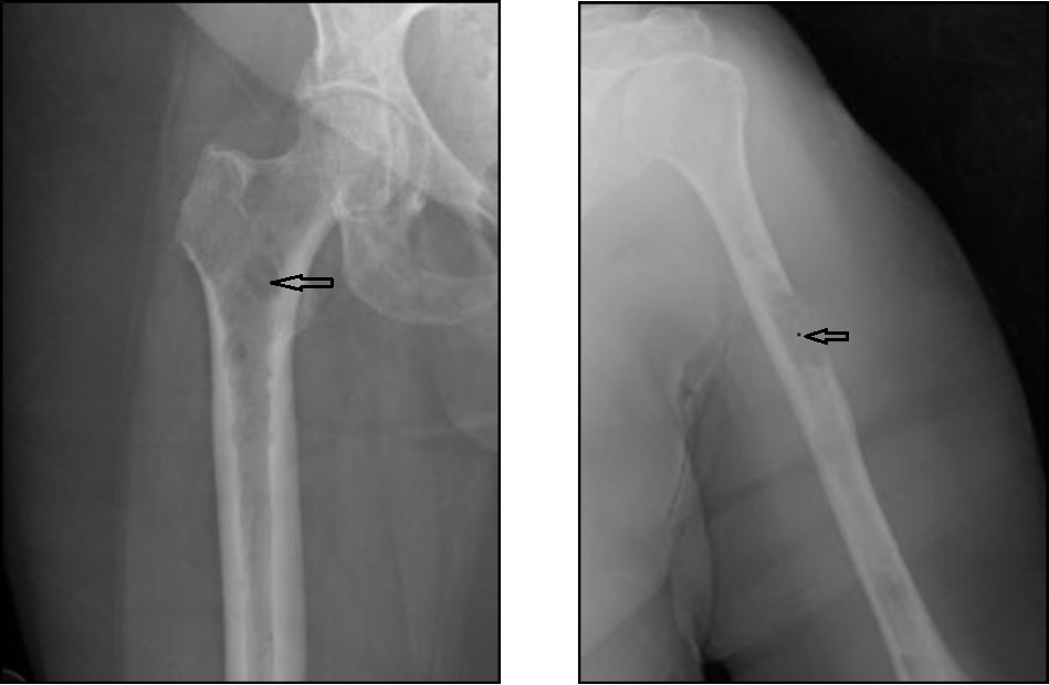
Figure 2.Several pathologic compression and burst fractures throughout the spine. Ventral cord indentation at T1, T8 and T11. Rostrocaudal cord edema spanning T10-T12 secondary to more significant cord compression. Bulky left paraspinal osseous/extraosseous disease at T8 (arrow) causing potential impingement.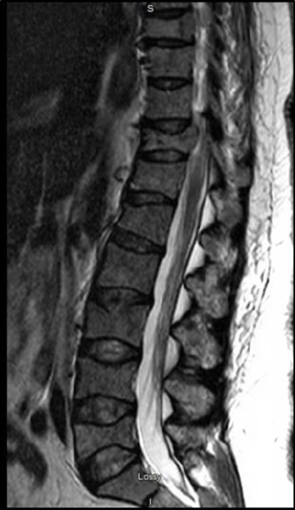
Discussion
Anion gap gets significantly altered depending on type of MM 5. Low AG is commonly observed in IgG myeloma due to its cationic property 5. In contrast, IgA myeloma can have an increased or a normal AG attributed to its anionic property 5. Pseudohyperphosphatemia, not a widely known phenomenon, can result from laboratory error 2,3. Paraproteinemias can also result in pseudohyponatremia due to the reduction in the plasma water fraction secondary to increased total protein concentration. Our case not only demonstrates an unusual presentation of MM with increased AG but also highlights the importance of recognizing paraproteinemias as a potential source of discrepant laboratory results before instituting inappropriate investigation and therapy for hyperphosphatemia or hyponatremia.
References
- 1.H G Drexler, Matsuo Y. (2000) Malignant hematopoietic cell lines: in vitro models for the study of multiple myeloma and plasma cellleukaemia. , Leuk. Res 24, 681-703.
- 2.J A Caras. (1997) Spurious hypophosphatemia associated with multiple myeloma. EndocrPract. 3, 135-136.
- 3.Mandry J M, Posner M R, Tucci J R, Eid C. (1991) Hyperphosphatemia in multiple myeloma due to phosphate-binding immunoglobulin. , Cancer 68(5), 1092-1094.