Juvenile Idiopathic Arthritis: A Study of 74 Cases in Northeast Brazil
Abstract
Objectives:
To identify the clinical epidemiological characteristics of patients with juvenile idiopathic arthritis (JIA) monitored at the Lucidio Portella Children’s Hospital (HILP) in northeastern Brazil and to ascertain the frequency and forms of presentation, the most affected joints, the most common joint involvement for each form, the frequencies of positive rheumatoid factor (RF+) and positive antinuclear antibodies (ANA+) in the various forms of presentation, and the most common complications.
Methods:
A study was conducted with 74 medical records of patients with JIA monitored at HILP between January 2010 and January 2013. Descriptive statistics were used for statistical analysis.
Results:
JIA was predominant in females with a mean age at onset of 5.2 years and a median disease duration of two years. The most frequent initial form of presentation was oligoarticular (63.5) arthritis, and the most affected joint was the knee (86.4%). The knee was most affected by oligoarticular arthritis, the wrist, knee, and ankle were affected by RF+ polyarticular arthritis, and the knee, ankle, and cervical spine were affected by systemic arthritis. RF+ was observed in 8.5% of the oligoarticular arthritis cases. ANA+ were present in 27.7% of the oligoarticular cases, in 22.2% of the systemic arthritis cases, and in 11.1% of the RF+ polyarticular arthritis . The most common complications were deformities (20.3%) and uveitis (14.9%).
Conclusion:
The findings for JIA patients in a referral hospital in northeastern Brazil were consistent with the literature regarding most of the evaluated criteria.
Author Contributions
Academic Editor: Kasapcopur Ozgur, Istanbul University
Checked for plagiarism: Yes
Review by: Single-blind
Copyright © 2017 Catarina fernandes pires, et al.
 This is an open-access article distributed under the terms of the Creative Commons Attribution License, which permits unrestricted use, distribution, and reproduction in any medium, provided the original author and source are credited.
Competing interests
The authors have declared that no competing interests exist.
Citation:
Introduction
The term juvenile idiopathic arthritis (JIA) represents a heterogeneous group of diseases characterized by arthritis in at least one joint with a minimum of six weeks duration and onset of symptoms up to 16 years of age1.It is not a single disease, but a group of related, genetically heterogeneous, phenotypically diverse immunoinflammatory disorders affecting joints and others structures, possibly activated by contact with an external antigen or antigens 2.
Chronic arthritis in children is not rare, but the true frequency is not known2. According to Ravelli and Martini, prevalence rates between 16 and 150/100,000 have been reported in the general population3.
It appears to be worldwide in distribution, but the reported incidence and prevalence vary considerably throughout the world4, 5. This fact may be reflected by the immunogenic susceptibility, ethnicity, and environmental influences of the populations studied 6, or may result from underreporting especially in developing countries where data are scarce and lack of unanimity in the choice of classification used in the various studies 2. Little is known regarding JIA in Brazil because epidemiological studies of JIA in Brazil are rare7.
Santos conducted a study in 72 patients in Belo Horizonte – Brazil, using the ILAR criteria and found the oligoarticular form as most frequent, 44.4% of cases7. Ramos, analyzed 100 children with JIA in São Paulo – Brazil, using the ACJ criteria and the most frequently found form was the oligoarticular 52% of cases8.
Oligoarticular form is the most frequent occurring in 50 to 60% of cases of JIA9, 10, is characterized by presence of chronic arthritis in up to four joints in the first six months of disease. According to ILAR criteria, the oligoarticular form is also classified as persistent (maintain up to four joints affected after six months of disease) or extended (affects five or more joints after initial six month period). There is a predominance of females with peak of incidence between one and tree years of age, although it may occur in children less than one year old and in adolescents2, 5, 9, 10, 11 .
Systemic Arthritis comprises 10% to 20% of JIA cases. About two-thirds occur in children under 5 years, although it can occur in any age. Genders are involved in same proportion, although there is a slight predominance in females when the onset occurs after 10 years of age12.
Polyarticular form onset corresponds 30 to 50% of patients with JIA and is characterized by chronic involvement of five or more joints during the first six months of disease. It includes forms with Negative Rheumatoid Factor (RF -) in 20% to 40% of JIA and with Positive Rheumatoid Factor (RF +) in 5% to 10% of JIA 2, 3, 10, 13, 14 . Classification is based in presence or absence of RF in serum of patients with JIA polyarticular form. RF is considered marker of disease and prognosis in JIA with this form onset, characterizing patients with severe erosive and limiting disease, higher frequency of subcutaneous nodules and vasculitis. The positivity of Rheumatoid Factor is associated with aggressive disease with joint deformities, reduction joint space, and erosion joint 15, 16.
Psoriatic arthritis is inflammatory seronegative arthritis for rheumatoid factor associated with cutaneous psoriasis. It is uncommon in childhood, accounting only 3% of chronic arthritis. Age of onset symptoms is usually between 9 and 12 years. It doesn’t usually have Gender predominance, except specific subtypes with predominance of females in polyarticular form symmetrical and males in spondylithic form17.
Undifferentiated Arthritis include diseases that some reason do not fills criteria for specific category or fills criteria for more than one 2, 10, 13, 14.
The diagnosis is o exclude other pathologies and identify subtypes of the disease associated with specific immunological and serological markers and to evaluate the degree of inflammation and systemic effects.10
According to Bisotto et al18, JIA patient care is based on a multidisciplinary model that includes physiotherapy, occupational therapy, and pharmaceutical therapy. Occasionally, surgery is required to correct deformities, and psychological support may also be required19.
The research objectives were to identify the clinical and epidemiological characteristics of JIA cases in their various forms of presentation, according to the classification of ILAR, and the most common complications, monitored in a Brazilian University Hospital.
Patients and Methods
The study was based on the medical records of 74 patients diagnosed with JIA according to the ILAR criteria (1997)10 who were monitored regularly in the Pediatric Rheumatology Service of HILP, which is a tertiary and teaching hospital associated with UFPI in Teresina, Piauí state (PI) in northeastern Brazil from January 2010 to January 2013. All the patients resided in the state of Piauí.
The following variables were studied: gender, ethnicity, age at presentation of the first signs/symptoms, disease duration, and form of initial presentation of the disease. The Caucasian and non-Caucasian classification was conducted in accordance with the phenotype of skin color informed by patient self-definition.
Data were also collected regarding the joints most affected, the most common joint involvement in every form, the frequency of RF+ in other forms of presentation (IgM RF+), and the identification of forms of JIA with ANA+.
RF was evaluated using the latex agglutination and Waaler-Rose tests with a value of 1:16. RF+ is associated with an aggressive disease progression including joint deformities, joint-space narrowing, and joint erosion20.
A continuous line of human tumor cells (HEP-2) were used to perform the ANA analysis. Titers between 1:80 and 1:160 were considered to be low, titers between 1:160 and 1:640 were considered to be medium, and titers above 1:640 were considered to be high. ANA is usually positive in low titers (less than 1:640) in 65% to 85% of children with oligoarticular JIA and in girls with uveitis21.
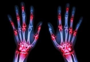
All the patients underwent a routine clinical rheumatologic examination. Complications included deformities, uveitis, and macrophage activation syndrome. The deformities are diagnostic for clinical manifestations in joints. The deformities that develop at the hands include ulnar drift at the wrists and the metacarpal phalangeal (MCP) joints and boutonniere and swan neck deformities at the fingers. The deformities that develop at the feet include hallux valgus deformities at the first MTP joints, hammertoe, and cock-up toe deformities. The deformities that develop at the knee include flexion contracture, epiphyseal enlargement, and hypoplasia of the menisci16.
All of the patients diagnosed with JIA underwent an ophthalmological slit lamp examination. Ocular involvement was considered when the ophthalmologist diagnosed uveitis based on the criteria established by the International Uveitis Study Group 3 adapted by the SUN2, according to the anatomical classification, considering previous, uveitis in which the primary site of inflammation was in the anterior chamber (iritis, anterior cyclites and iridocyclites)22. Another criterion adopted for the classification of uveitis was clinical manifestations such as hyperemia, eye pain, tearing, decreased visual acuity, photophobia, and headache.
Macrophage activation syndrome, which is the most serious complication of systemic JIA, was diagnosed using a symptomatology of sudden onset of fever, generalized lymphadenopathy, hepatosplenomegaly, and coagulation disorders and was confirmed by the presence of histiocytic phagocytosing hemocytes or platelets in the bone marrow, lymph nodes, liver, or spleen, low ESR, and pancytopenia.
Statistical Analysis
Data were collected on a sheet, tabulated, and processed using the Statistical Package for the Social Sciences 17.0 (SPSS) program, and the results are presented in tables and graphs.
For the statistical analysis, frequencies were calculated for the categorical qualitative variables. For the quantitative variables, the central tendency was determined using the mean and median, and the dispersion was determined using the standard deviation.
Results
Of the total 74 patients studied, 47 (63.5 %) were female and 27 (36.5%) were male, 51 were non-Caucasian (69.2%), and 23 were Caucasian (38.8%).
The mean age at presentation of the first signs/symptoms was 5.2 years (SD = 3.3 years) with a minimum age at the first signs/symptoms of one year and a maximum of 12 years.
The median disease duration was two years, the minimum duration was six months, and the maximum duration was 11 years.
Oligoarticular arthritis represented the most frequent presentation (47; 63.5%) followed by systemic (11; 14.8%) and RF+ polyarticular (9; 12.2%) arthritis. RF- polyarticular (4; 5.4%) and psoriatic arthritis were present in three cases (4.1%). There were no cases related to enthesitis-related arthritis or undifferentiated arthritis (Table 1).
Table 1. Epidemiological characteristics of patients with JIA in the various forms of the disease in Brazilian Piauiense Children (n=74)| Initial | Sex | Age onset (year) | Disease duration (year) | |||
| form n % | Male Female n % n % | Mean (Sd) Median | Mean(Sd) Median | |||
| Oligo (47; 63.5) | 15 31.9 | 32 68.1 | 4 (±3) | 3 | 3 (±2) 2 | |
| Systemic (11; 14.8) | 6 54.5 | 5 45.5 | 6 (±2) | 7 | - | 0.5 |
| RF+ Poly (9; 12.2 ) | 1 11.1 | 8 8.9 | 7 (±3) | 8 | 5 (±3) | 1 |
| RF- Poly (4; 5.4) | 4 100 | - - | 7 (±4) | 8 | 1 (±0.5) | 0.5 |
| Psoriatic (3; 4,1) | 1 33.3 | 2 66.7 | 11 (±4) | 9 | - | 2 |
Oligoarticular arthritis was most commonly observed in the knee. RF+ polyarticular arthritis was frequently observed in the wrist, knee, and ankle. RF- polyarticular arthritis was observed to the same extent in the knee, ankle, wrist, and cervical spine. The systemic form most frequently involved the knee and ankle joints followed by the cervical spine. The knee joint was involved in most of the psoriatic arthritis cases (Table 2)
Table 2. Distribution cases with Juvenile Idiopathic Arthritis accompanied in HILP from January 2010 to January 2013, due to presentation of disease and type affected joint (n=74).| Forms | OOligoart ((n=47) | Polyartic.FR+ (n=9) | Polyartic.FR- (n=4 ) | Systemic Psorias Total (n=11) (n=3) (n=74) | |
| affected joint | n % | n % | n % | n % n % n % | |
| Cervical spine | - - | 5 55.5 | 1 25.0 | 7 63,3 - - 13 17.6 | |
| Mandibular | - - | - - | - - | 3 27.3 - - 3 4.0 | |
| temporo | |||||
| Shoulder | - - | - - | - - | 1 9.0 - - 1 1.5 | |
| Elbow | 2 5.5 | 5 55.5 | - - | 4 36.5 - - 11 14.8 | |
| Wrist | 5 11.1 | 9 100.0 | 3 75.0 | 6 54.5 1 33.3 24 32.4 | |
| Hand | 4 8.3 | 7 77.7 | 1 25.0 | 1 9.0 1 33.3 14 18.9 | |
| Coxo-femoral | 2 5.5 | 1 11,1 | 1 25.0 | 4 36.5 - - 8 10.8 | |
| Knee | 40 86,1 | 9 100,0 | 4 100.0 | 8 72.7 3 100.0 64 86.4 | |
| Ankle | 13 27.7 | 9 100.0 | 3 75.0 | 8 72.7 1 33.3 34 45.9 | |
| Foot | - - | 3 33.3 | - - | - - - - 3 4.0 |
The frequency of serological characteristics and most commonly observed complications are shown in Table 3.
Table 3. Distribution frequency of serological characteristics and main complications JIA patients followed in HILP from January 2010 to January 2013 (n = 74).| Disease Forms | O Oligoart ((n=47) n % | Polyartic.FR+ (n=9) n % | Polyartic.FR- (n=4 ) n % | Systemic Psorias Total (n=11) n% (n=3) n% (n=74) n% |
| Serological characteristics | ||||
| FR+ | 4 8.5 | 9 100.0 | - - | - - - - 13 17.5 |
| FAN+ | 16 34,0 | 2 22,2 | - - | 2 18,2 - - 20 27,0 |
| Complicações | ||||
| Deformities | 9 19.1 | 6 66.6 | - - | - - - - 15 20.3 |
| Uveitis | 11 23,4 | - - | - - | - - - - 11 14,9 |
| Macrophage | - - | - - | - - | 1 9.1 - - 1 1,3 |
| activation | ||||
| syndrome |
Discussion
This study showed that JIA most commonly affects female non-Caucasian children. Santos7 studied 72 patients with JIA in the state of Belo Horizonte – Minas Gerais and found no significant difference between the two genders in the sample.
A higher frequency of JIA was observed in non-Caucasian patients. The classification was performed regarding the self-defined phenotype of skin color. Brazil represents a mixed population with a relatively large percentage of immigrants, especially in the Northeast, which prevents an exact determination of patient ethnicity difficult.
In a multicenter study conducted in 13 Brazilian pediatric rheumatology referral centers, 780 children with JIA were treated during a one-year period. In the Pediatric Rheumatology Outpatient Unit of the Department of Childcare and Pediatrics, School of Medicine of Ribeirão Preto - Brazil, an average of 14 new cases of JIA per year were treated over the past five years23. The minimum age of onset of signs / symptoms occurred precocious (Table 1), which is worrying, considering that as children begin to walk around one year of age and are already affected by disease.
Two peaks of a higher incidence of disease onset were determined: children under five, who belong predominantly to the oligoarticular group, and adolescents, who belong to the RF+ and enthesitis-related arthritis groups24 (Table 1).
The most frequently affected joint was the knee followed by the heel, wrist, hand, and cervical spine (Table 2).
Oligoarticular arthritis (63.5%) was the most frequently observed initial presentation of JIA in the sample, which is consistent with the literature. In Europe and North America, oligoarticular arthritis is the most frequent form of JIA and represented approximately 50% of the cases in a large case study series conducted in 1997 by Chalom et al25. RF+ arthritis is less frequent (Table 4). There is considerable variation in the relative proportions of the other forms. In Asia, oligoarticular arthritis is the most frequent disease presentation9 and is also the most frequently observed form in Turkey, France, Spain, the UK, South Africa, and Italy2, 17, 26, 27, 28, 29. Systemic arthritis (14.7%) was the second most frequent disease presentation, which was consistent with the findings presented by Santos et al7 but inconsistent with worldwide literature ranking systemic arthritis as the third most common disease presentation. The finding in the present sample can be attributed to more judicious investigation and diagnosis because systemic arthritis demonstrates the most acute manifestations and frequently requires the patient to access health services including hospitalization.
Table 4. Comparative epidemiological data for Juvenile Idiopathic Arthritis (%)| Forms | Piauí Brazil Pires et al, 2014 | BeloHorizonte Brazil Santos, 2006 | Turkey Yilmazet al, 2008 | India Kunjir et al, 2010 | South of África, Weakley et al, 2012 | United Kingdom Thomson et al, 2002 | Spain Merino et al, 2001 | Alsace, France Danner et al, 2006 | Italy Ravelli, 2007 |
| Systemic | 14,9 | 31,9 | 15,3 | 8,0 | 7,7 | 14,5 | 14,4 | 8,9 | 4 - 17 |
| Oligo | 63,5 | 34,7 | 34,1 | 21,0 | 26,8 | 45,6 | 37,6 | 40,3 | 27 -56 |
| Poly FR- | 5,4 | 18,1 | 30,6 | 17,0 | 26,9 | 19,6 | 22,4 | 20,4 | 28-Nov |
| Poly FR+ | 12,2 | 4,2 | 6,6 | 12,0 | 14,0 | 7,1 | 1,6 | - | 2 - 7 |
| Psoriat A | 4,0 | 4,2 | 1,0 | 1,0 | 1,3 | 7,1 | 1,6 | 4,5 | 11-Feb |
| ERA | _ | 5,6 | 10,3 | 36,0 | 23,0 | 6,5 | 7,2 | 17,9 | 11-Mar |
| others | _ | 1,4 | 2,5 | 5,0 | - | - | 15,2 | 6,0 | 21-Nov |
RF+ polyarticular arthritis was the third most frequently observed disease presentation with 9 cases (12.2%), which is also higher than values reported in the literature (Table 4). Similar results were found in India and southern Africa (Table 4) The great majority of European countries have observed RF+ polyarticular arthritis is around 7% among all JIA forms; in Spain, the frequency is low, around 1.6%(Table 4). RF+ polyarticular arthritis was higher our research due to most necessity of patients in seeking the health services by being a severe disease with clinical phenotypic, Serological and Immunogenetic similarities to Adult Rheumatoid Arthritis. Severe and chronic joint impairment in child is capable of causing inability to perform normal activities and if undiagnosed and treated in a hard time is able to cause sequelae definitively as joint deformities. The four cases (5,4%) of RF- polyarticular arthritis in the present study represented a lower frequency compared with the literature, which commonly determines RF- arthritis to be the second most common disease presentation. The clinical manifestations of RF- arthritis are typically less painful, which may cause patients to delay seeking health services or remain undiagnosed. A recent study showed that joint swelling and gait disorders were the most frequent causes of referral of children who were later diagnosed with JIA23. A larger study sample may have included a higher proportion of RF- cases more similar to the literature.
Psoriatic arthritis was found in a small number of cases (3; 4.0%) and more often in females. Skin lesions were manifested before joint involvement. In male patients, skin involvement occurred at the same time as joint involvement and was severe. Psoriatic arthritis was both RF- and ANA-. Statistics have shown that 6-42% of psoriasis patients develop some type of joint involvement17. Psoriatic arthritis does not usually exhibit gender predominance, except in specific subtypes; females more commonly exhibit the symmetrical polyarticular form, and males more commonly exhibit the spondylitic form. Skin involvement usually precedes arthritis in 75% of cases with simultaneous onset in 10% of patients. In the remaining 15%, arthritis can occur after skin injury. It is unusual to find a correlation between the type or severity of skin lesions and the presence, type, or extent of joint involvement17, 30.
Enthesitis-associated arthritis and undifferentiated arthritis, which are commonly under-diagnosed and exhibit unspecific forms of clinical presentation, were not detected in this study (Table 4)
The most commonly affected joint was the knee (86.4%), which is inconsistent with the literature, followed by the ankle (45.9%), wrist (32.4%), hand (18.9%), cervical spine (17.6%), elbow (14.8%), and coxofemoral joint (10.8%). Arthritis can involve any joint. The initial phase most frequently involves oligoarticular arthritis, which can progress to polyarthritis24.In the present study, the joints most commonly affected by oligoarticular arthritis were similar to previous studies: the knee was affected in 86.1% of cases followed by the ankle in 27.7% of cases. The study by Huemer et al31 with 64 children with oligoarticular JIA, the knee joint was the most affected joint in the initial form of the disease (89%) followed by the ankle joint (36%).
The study revealed that the knee and ankle were equally affected in the systemic form of arthritis (72.7% of cases). The joints of the cervical spine and wrist were affected more than half the cases. According to Petty and Cassidy12, joint involvement in systemic arthritis may be minimal early in the disease and become severe over weeks or months. Several joints may be affected at the beginning or during the course of the disease. Monoarticular involvement is uncommon, and development is typically polyarticular. The knee, wrist, and ankle are the most commonly involved joints. Involvement of the cervical spine, hip, and temporomandibular joint occurs in over half of patients with the systemic form of JIA12.
The wrist, knee, ankle and metacarpophalangeal joints were most affected by RF+ polyarticular arthritis, which is consistent with previous studies. In RF+ polyarticular arthritis, the small and large joints of the upper and lower extremities are affected to the greatest extent along with the cervical spine and temporomandibular joint. The characteristic pattern is symmetrical arthritis, which mainly affects the metacarpophalangeal, metatarsophalangeal, and wrist joints.1
The knee, wrist and ankle spine joints were most affected in cases of RF- polyarticular arthritis. According to Petty and Cassidy2, the knee, wrist, and ankle joints are the most commonly affected joints among children with RF- polyarticular arthritis in the initial and later stages.
RF+ arthritis was present in 17.5% of the surveyed sample, in all the cases of RF+ polyarticular arthritis, and in 8.5% of the oligoarticular arthritis cases. RF+ has been shown to occur in 7-10% of children with JIA using latex agglutination and Waaler-Rose tests. Using an enzyme-linked immunosorbent assay (ELISA), the values increase to 22%-35%. RF+ is usually present in female children with polyarticular JIA and indicates a severe and erosive disease2 and a worse prognosis. Children with RF+ polyarticular JIA should undergo stricter monitoring to prevent joint sequelae.
ANA + were present in 27.0% of the total sample surveyed, in 34.0% of cases of the oligoarticular arthritis cases, 18.2% of the systemic arthritis cases, and 22.2% of the RF + polyarticular arthritis cases. Probably, difference between results found in the study and other studies is due to number of the sample searched. Possibly, the expansion the studied population may show results closer to those in literature. In Brazil, researches between FAN and JIA are strongly associated with the presence of uveitis. Previous studies have shown that ANA are usually positive in 40% to 85% of cases with early oligoarticular arthritis. Several researches have revealed strong association between positive ANA with oligoarticular JIA and uveitis in a female child 28, 32. AN is positive in several autoimmune rheumatic diseases33, 34. In JIA, FAN usually has a low to moderate titre.
Deformities were observed in 20.3% of the cases and were distributed in 19.1% of the oligoarticular arthritis cases and 66.6% of the RF+ polyarticular arthritis cases. RF+ polyarticular arthritis presents several clinical, immunogenic, and developmental similarities with rheumatoid arthritis (RA) in adults. Synovitis persisting for months or years may cause irreversible consequences such as subluxations, fusions, or osteoarticular destruction and may evolve into functional incapacity with important implications for the quality of life of individuals and their families35.
Uveitis was observed in 14.9% of the cases. The frequency of chronic uveitis has varied considerably in a series of reports of children with chronic arthritis: a rate of 2% was found in Costa Rica36, 10% in the United States, 13% in Canada25, and 16% in the Nordic countries37.
Uveitis exhibits an insidious and asymptomatic onset in most cases. Untreated uveitis can evolve with sequels ranging from posterior synechiae to cataracts, glaucoma, and blindness38, especially in children with oligoarticular onset JIA with ANA+.
Macrophage activation syndrome occurred in only one case (1.3%) in the present study. This is the most serious complication of systemic JIA. It is considered a form of hemophagocytic lymphohistiocytic syndrome and is associated with serious morbidity. Macrophage activation syndrome occurs in 7% of cases during the course of the disease. Early recognition and appropriate treatment of this complication are essential for survival of the patient39.
This study draws attention to the early age of onset, prolonged disease duration, affected joints, and severe and disabling complications of JIA in a phase of life where health is essential for proper growth and development in northeastern Brazil. The importance of early diagnosis, proper treatment, and regular monitoring with free access to health services must be emphasized. Due to the importance of the topic, the scarcity of Brazilian scientific studies on the topic, and the serious consequences for the health of children, further studies of this disease are necessary.
References
- 1.Weldt L L, Aguilera M M, Loyola M T. (2001) . Pediatric Rheumatology. 2th ed. Rio de Janeiro:Revinter .
- 6.Manners P J, Bower C. (2002) Worldwide prevalence of juvenile arthritis-why does it vary so much?. , J Rheumatol 29, 1520-30.
- 7.Oen K. (2000) Comparative epidemiology of the rheumatic diseases in children. Curr Opin Rheum. 12, 410-14.
- 8.Anderson G B. (1999) Juvenile arthritis – who gets it, where and when, a review of current data on incidence and prevalence. , Clin. Exp. Rheumatol 17, 367-374.
- 9.FPTS Santos, MAP Carvalho, Pinto J A, ACH Rocha, Campos W R. (2010) Juvenile Idiopathic in a Department of Rheumatology: Belo Horizonte, Minas Gerais. Rev Med Minas Gerais. 20(1), 48-53.
- 10.VCSR Ramos, Ronclezel M V, Okuda E M, Sacchetti S B. (2006) Caracterização Epidemiológica, clínica e laboratorial de 100 crianças com artrite reumatoide juvenil. Rev Paul Pediatria. 24(4), 335-42.
- 11.Oen K G, Cheang M. (1996) Epidemiology of chronic arthritis in childhood. Seminars in arthritis and rheumatism. 26, 575-91.
- 12.Petty R E, Southwood T R, Baum J, Bhettay E, Glass D N et al. (1997) Revision of the proposed classification criteria for juvenile idiopathic arthritis. , Durban;, J Rheumatol 25, 1991-94.
- 13.Manners P J, Bower C. (2002) Worldwide prevalence of juvenile arthritis-why does it vary so much?. , J Rheumatol 29, 1520-30.
- 14.Benedetti F DE, Schneider R. (2011) . Systemic Juvenile Arthritis. In:Cassidy,JT, Petty,RE, Laxer,RM, Lindsley CB.Textbook of Pediatric Rheumatology.6a ed.Philadelphia:WB Saunders; (Section 2-Chapter14) 236-248.
- 15.Petty R E, Southwood T R, Baum J, Bhettay E, Glass D N et al. (2001) International League of Associations for Rheumatology classification of juvenile idiopathic arthritis, second revision. , Edmonton 31, 390-92.
- 16.Weld L L, Aguilera M M, Loyola M T. (2001) Artrite Idiopática Juvenil. In:. Oliveira SKF & Azevedo ECL. Reumatologia Pediátrica. 2 Ed. Rio de Janeiro Revinter; 143-208.
- 17.Ferreira R A, Silva C H, Silva D A, Sopelete M C, Kiss M H et al. (2007) Is measurement of IgM and IgA rheumatoid factors (RF) in juvenile rheumatoid arthritis clinically useful? Rheumatol Int. 27, 345-9.
- 19.Lindsley C B. (2011) Textbook of Pediatric Rheumatology. 6a ed. Philadelphia: WB Saunders.(Section 2-Chapter 15) 249-61.
- 20.Espinosa L R, Cuellar M L. (1998) Psoriatic arthritis and spondylitis: a clinical approach. In: Calin A, Taurog JD (editors). Spondylarthritides , Oxford: OxfordUniversityPress 97-111.
- 21.Bisotto L S, Xavier R M, Machado S H, Bredmeier M, JCT Brenol. (2005) Impact of inflammatory activity and glucocorticoid use in the nutritional variables of juvenile idiopathic arthritis. Rev Bras Rheumatol, São Paulo. 45-5.
- 22.Bueno V C, Lombardi I, Medeiros W M, MMA Azevedo, Len C A et al. (2007) Rehabilitation in Juvenile Idiopathic Arthritis. Rev Bras de Rheumatol, São Paulo. 47(3), 197-203.
- 23.Ferreira R A, Silva C H, Silva D A, Sopelete M C, Kiss M H et al. (2007) Is measurement of IgM and IgA rheumatoid factors (RF) in juvenile rheumatoid arthritis clinically useful? Rheumatol Int. 27, 345-9.
- 24.Petty R E, Cassidy J T, Sullivan B D. (1973) Clinical correlates of antinuclear in juvenile rheumatoid arthritis. , J Pediatrics 83, 386-389.
- 25.Jabs D A, Nussenblatt R B, Rosenbaum J T. (2005) Standardization of Uveitis Nomenclature (SUN) Working Group. Standardization of uveits nomenclature for reporting clinical data. , Results of the First International Workshop.Am J Ophthalmol.Review 140(3), 509-16.
- 26.MFF Carvalho, Magalhães C, Silva C A, Sztanjnbok F, MOE Hilario et al. (2005) Epidemiology of pediatric rheumatic diseases in Brazil: multicentric study. Clin Exp Rheumatol. 23-76.
- 27.Sztajnbok F R, CRB Serra, MCF Rodrigues, Mendonza E. (2001) Rheumatic diseases in Adolescence. , J Pediatri, Rio Janeiro 77, 234-244.
- 28.Chalom E C, Goldsmith D P, Koehler M A, Bittar B, Rose C D et al. (1997) Prevalence and outcome of uveítes in regional cohort of patients with juvenile rheumatoid arthritis. , J Rheumatol 24, 2031-2034.
- 29.Yilmaz M, Kendirli G, Altintas D U, Karakoc G B, Inal A et al. (2008) Juvenile idiopathic arthritis profile in turkish children. Pediatr Int. 50, 154-158.
- 30.Merino R, J De Inocencio, Garcia-Consuegra J.(Dec2001) Evatoluation of ILAR criteria for juvenile idiopathic arthritis in Spanish children. , J Rheumatol 28(12), 2731-6.
- 31.Thomson W, Barrett H, Donn R, Pepper L, Kennedy L J et al. (2002) Juvenile Idiopathic arthritis classified by the ILAR criteria: HLA in UK patients. Rheumatology. 41, 1183-89.
- 32.Weakley K, Esser M, Scott C. (2012) Juvenile idiopathic arthritis in two tertiary centres in the Western Cape, South Africa. Pediatr Rheumatol. 10-35.
- 33.Sampaio-Barros P D, Azevedo V F, Campos W R, Carneiro SCS, Carvalho MAP et al. (2007) Brazilian Consensus on Spondyloarthropathies: ankylosing spondylitis and psoriatic arthritis diagnosis and treatment - first review. Rev Bras de Rheumatol, São Paulo:. 47, 234-243.
- 34.Huemer C, Mallsenon P N, Cabral D A, Huemer M, Falger J et al. (2002) Patterns of joint involvement at onset differentiate oligoarticular juvenile psoriatic arthritis from pauciarticular juvenile rheumatoid arthritis. , J Rheumatol 29, 1531-1535.
- 35.Ploski R. (1997) Immunogenetic polymorphism and disease mechanisms in juvenile chronic arthritis. Rev Rhum. , 64 (Suppl) 10, 127-130.
- 36.Silva N P, LEC Andrade. (2006) Laboratório em reumatologia. In: Sato E. Guias de medicina ambulatorial e hospitalar da Unifesp /Escola Paulista de Medicina. , Reumatologia. Barueri: Manole 17-34.
- 37.Kiss M H, Silva CHM, Ferriani V M. (2003) Exames laboratoriais em reumatologia pediátrica. Laboratório em reumatologia pediátrica. In:Marcondes E, Vaz FCA,Ramos JLA, Okay Y. Pediatria Básica.Tomo II.Pediatria clínica e geral.9.Ed.São Paulo:Sarvier. 777-81.
- 38.Bueno V C, Lombardi I, Medeiros W M, MMA Azevedo, Len C A et al. (2007) Rehabilitation in Juvenile Idiopathic Arthritis. Rev Bras de Rheumatol, São Paulo. 47(3), 197-203.
- 39.Arguedas P, Fasth A, Andersson-Gare B. (2002) A prospective population based study of outcomes of juvenile chronic arthritis in Costa Rica. , J Rheumatol 9, 174-183.
- 40.Kunnamo I, Kallio P, Pelkonen P. (1986) Incidence of arthritis in Urban Finnish children: a prospective study. Arthritis Rheum. 29, 232-238.